Int J Cardiovasc Imaging
Presence of aortic root vortex formation after TAVI with CENTERA confirmed using 4D-flow magnetic resonance imaging
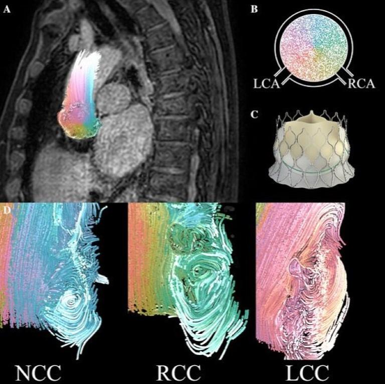
Novel nitinol, self-expandable, short bodied transcatheter aortic valve implantation (TAVI) prostheses allow for magnetic resonance imaging (MRI)-based evaluation of the left ventricle and aortic root, due to a relatively small susceptibility artefact. We have employed four dimensional (4D-)flow MRI-analysis in 3 patients, 2 years after uncomplicated TAVI with the new CENTERA prosthesis. Thoracic 4D flow MRI was conducted at 3.0T with a spatial and temporal resolution of 2.5 mm3 and ± 42 milliseconds. Vortex formation in the three sinuses of Valsalva was analyzed using advanced streamline techniques.
More Information
Citation
BibTex
@article{bib:vendrik:2018,
    author       = { Vendrik, Jeroen and Farag, Emile and de Hoon, Niels and Kluin, Jolanda and Baan, Jan },
    title        = { Presence of aortic root vortex formation after TAVI with CENTERA confirmed using 4D-flow magnetic resonance imaging },
    journal      = { Int J Cardiovasc Imaging },
    volume       = { 34 },
    year         = { 2018 },
    pages        = { 1947--1948 },
    doi          = { 10.1007/s10554-018-1413-2 },
    pubmedid     = { 30094565 },
    url          = { https://publications.graphics.tudelft.nl/papers/204 },
}